OCD definition
OCD, also known as osteochondrosis/osteochondritis dissecans, is a disease of the developing joints that is most often seen in young, growing patients. In addition to humans, this condition has been described in several domestic animal species, including dogs, horses, and pigs. The disorder is characterized by the formation of cracks, flaps, and/or loose bodies, composed of cartilage and underlying bone, within the affected joints. OCD lesions in humans most often develop in the knee joint and less frequently involve the elbow and ankle joints.

In this handout we will provide you with MRI and histology images that illustrate current thoughts about the most likely pathway for the development of OCD and its progression to clinical disease.

How does OCD develop?
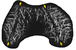
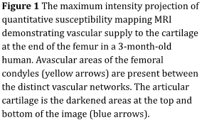
The details of the development of OCD remain incompletely understood, but recent studies performed in human and animal patients indicate that the development of OCD lesions starts well before the appearance of clinical signs, such as pain. Early in development, the ends of the growing long bones are composed entirely of cartilage. The surface articular cartilage, which provides the smooth gliding surface of the tissue, obtains its nutrition from the synovial fluid. Conversely, the remainder of the end of the growing long bone is composed of epiphyseal (also known as “growth”) cartilage which requires a blood supply to maintain its viability (Figure 1). During growth, the epiphyseal cartilage is gradually replaced by bone in a process known as endochondral ossification. In adults, the only cartilage remaining is the surface articular cartilage. It is believed that the development of OCD starts with a disturbance of the blood supply to the epiphyseal growth cartilage which results in an area of the epiphyseal growth cartilage dying. This prevents replacement of the epiphyseal growth cartilage with bone via endochondral ossification.

What happens after the damage to the epiphyseal growth cartilage?
After damage occurs to the epiphyseal growth cartilage, these early lesions of dead growth cartilage may either heal or progress to clinical disease. Although it is not yet possible to predict which lesions will heal and which will not, it is thought that the majority of cases remain asymptomatic and go on to heal, as long as the overlying articular cartilage is not disrupted. The reparative process in small lesions may include surrounding of the area of dead cartilage by bone with gradual resorption of dead cartilage and replacement with bone (Figure 2). Larger lesions may have a more complex healing response including initial removal of dead cells and tissue by inflammatory cells, followed by replacement by fibrous tissue and blood vessels. There may also be extensive myelofibrosis (scar formation in the bone marrow cavity) and remodeling of the bone next to the lesion (Figure 2). In non-healing lesions, the articular cartilage appears thicker, because this tissue layer now includes the normal surface articular cartilage as well as an underlying zone of dead epiphyseal cartilage. (Figure 2). In some cases, areas of dead epiphyseal cartilage, that are not fully converted to bone, collapse because they are unable to support the overlying articular cartilage. This is especially the case if the articular cartilage has to sustain strong forces, such as in the case of sporting activities. Repeated exposure of these unsupported segments of articular cartilage to excessive forces may result in the development of cracks, flaps, and fragments extending through the articular cartilage, causing the clinical signs of OCD such as joint pain, locking and swelling. Once a lesion has advanced to this point, it will often require surgical treatment to promote healing.

Figure 2

  • Focal necrosis of the epiphyseal cartilage—caused by damage to the vessels supplying this area—resulting in the failure of the endochondral ossification which appears as an area of thickened cartilage (circled).
  • Small focus of retained damaged cartilage (black arrows) that has been surrounded by bone (pink material) and bone marrow (open spaces) without significant lasting disruption of bone formation.
  • Area of healing in a larger lesion, with early scar formation that includes proliferation of small blood vessels (black arrows) and fibrous tissue.

How does OCD present and how is it diagnosed?
Patients with OCD present with nonspecific symptoms of pain and sometimes locking in the joint. Symptoms evolve slowly and usually there is not one “event” that can be identified to have caused the symptoms. Patients are generally physically active, and the symptoms are often first noted during physical activities.

Evaluation of patients usually starts with obtaining an x-ray. The x-ray often shows an oval shaped abnormality in the bone just above the joint line. When a radiologist describes the OCD lesion, they often mention the parent bone, the progeny lesion, and the interface. The parent bone is the bone deep and adjacent to the lesion. The progeny lesion is the OCD lesion which can be composed of cartilage and bone, or cartilage only. The interface is the space in between the parent bone and the progeny lesion. X-rays are only able to detect the bony structures of joints, while all the soft tissues, including the cartilage, are invisible. When an injury occurs during the process of normal bone maturation, the parent bone has a concave appearance on an x-ray because the progeny lesion is not visualized as densely as the parent bone (Figure 3). Therefore, once there is a bony abnormality noted on x-ray, an MRI is the most appropriate next imaging modality because it will reveal changes in non-bony tissues as well.

Figure 3
AP and lateral radiograph of a 17-year-old male demonstrating healing of an OCD lesion (between arrows) at first visit (a) and after 8 months of non-surgical treatment (b). The lesion becomes smaller because more bone has been laid down which bridges between the progeny lesion and underlying parent bone.

How does OCD heal?
If the joint is protected from excessive loads and the articular cartilage is preserved, healing of the areas of dead growth cartilage is possible in young individuals through incremental replacement with bone. About 60% of OCD lesions successfully heal with 6 to 18 months of conservative, nonsurgical treatment, which typically involves sports restriction, casting, or bracing to decrease the load on the joint. The process of lesion healing starts in a region of the lesion adjacent to healthy subchondral bone and involves gradual replacement of cartilaginous and fibrous tissue by bone (Figure 2), followed by eventual integration into the subchondral bone (Figure 4). On MRI, it is possible to follow the progression of lesion ossification and bony bridging between the lesion and parent bone. For lesions that do not heal on their own, there is a variety of surgical interventions that can be utilized to produce bony healing.

Figure 4
Proton density-weighted MRI of the knee joint of an 11-year-old female demonstrating healing of an OCD lesion (between arrows) at first visit (a) and after 8 months of non-surgical treatment (b). The lesion becomes smaller because more bone has been laid down which bridges between the progeny lesion and underlying parent bone.

Summary
Although the pathogenesis is not yet completely understood, current knowledge indicates that, in most patients, the initial event occurring in OCD is an injury to blood vessels supplying the epiphyseal growth cartilage. This results in the epiphyseal growth cartilage dying and a focal failure of the normal endochondral ossification process (Figure 5). Secondary ossification of the lesion starts circumferentially (Figure 6) and later fills the inner portion of the lesion (Figure 7) with varying degrees of associated bridging to the underlying parent bone. Lesions that heal will completely bridge with the underlying bone and a linear “scar” will be all that remains (Figure 8). It is believed that the great majority of these lesions heal uneventfully. Conversely, in lesions that persist, the progeny lesion separates from the parent bone, resulting in a fluid filled gap that involves the articular cartilage. At that point, the lesion fragment is unstable and can produce symptoms either with the progeny fragment in place or after displacement of the fragment, which may cause symptoms in other parts of the joint.